
.jpg)
Pulp Therapy
Tooth Fairy's Vault: Exploring the Post-Thaw Stability of Dental Pulp Stem Cells After Long-Term Cryopreservation
 photo")
Malika A. Malik, DDS (she/her/hers)
Pediatric Dentistry Resident
University of Michigan, Ann Arbor, MI
University of Michigan
Ann Arbor, Michigan, United States
Alyssa Nedell, NA
University of Michigan
Elizabeth Perkins, NA
University of Michigan
Jim Sugai, NA
University of Michigan
Darnell Kaigler, n/a
University of Michigan
Ann Arbor, Michigan, United States
James R. Boynton, DDS, MS
Professor
University of Michigan- Ann Arbor
Ann Arbor, Michigan, United States
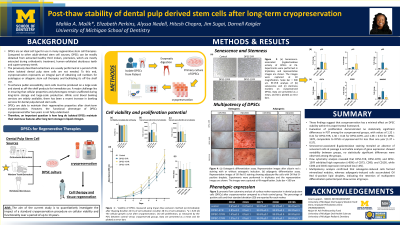
Background: There has been increased interest in the prospects of using dental pulp-derived stem cells (DPSCs) in clinical cell therapies due to their regenerative and immunomodulatory potential. DPSCs are relatively easy to harvest and can be cryopreserved for future therapeutic use, however, further analysis is required to assess the viability and stemness capacity of DPSCs following long-term cryopreservation. This study aimed to evaluate the viability and stem cell properties of DPSCs cryopreserved for up to 13 years.
Methods: DPSCs were cryopreserved for 5 (DPSC-5YR),10 (DPSC-10YR), and 13 (DPSC-13YR) years. Following thawing and early-passage culture, DPSCs were characterized via flow cytometry for their expression of cell surface markers (CD34, CD45, CD73, CD90, and CD105). Cell viability and total cell count were assessed via the trypan blue exclusion method, while proliferation capacity was evaluated via a 7-day Cell Counting Kit-8 (CCK-8) assay, and population doubling time (PDT) was determined. Multipotency was examined through differentiation staining, and gene expression analysis of stem cell- and senescence-related markers was conducted using reverse transcription-quantitative polymerase chain reaction (RT-qPCR). Additionally, senescence-associated β-galactosidase activity was assessed.
Results: Flow cytometry analysis revealed that DPSC-5YR, DPSC-10YR, and DPSC-13YR exhibited high expression (>90%) of CD73, CD90, and CD105, while CD34 and CD45 expression remained low ( <4%). Evaluation of proliferation demonstrated no statistically significant differences in PDT among the cryopreserved groups, with values of 1.32 ± 0.41 for DPSC-5YR, 1.36 ± 0.44 for DPSC-10YR, and 1.38 ± 0.53 for DPSC-13YR, comparable to DPSCs cryopreserved for less than one year (1.37 ± 0.57). Multipotency analysis confirmed that osteogenic-induced cells formed mineralized nodules, whereas adipogenic-induced cells accumulated Oil Red O-positive lipid droplets, indicating the retention of multipotent differentiation potential post-thaw across all groups. Senescence-associated β-galactosidase staining revealed an absence of senescent cells at passage 6 and while analysis of gene expression showed variability between groups, no statistically significant differences were observed among the groups.
Conclusions: This study is the first to demonstrate that DPSCs retain viability, proliferative capacity, stemness, and multipotency after 13 years of cryopreservation. These findings have significant implications for the clinical use of banked DPSCs for therapeutic applications of tissue regeneration and immunomodulation.