
.jpg)
Oral Pathology
A Case of Double Mesiodens in a 7-year-old Hispanic Male

David Neal, DMD
Pediatric Dental Resident, PGY-1
Indiana University School of Dentistry - Riley Hospital for Children
Indiana University School of Dentistry
Carmel, Indiana, United States
LaQuia A. Walker-Vinson, DDS, MPH, FAAPD
Associate Professor
Indiana University School of Dentistry / Riley Hospital for Children
Indianapolis, Indiana, United States
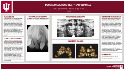
Case Report: This presentation discusses a 7-year-old male patient who presented to comprehensive chldren’s hospital outpatient dental clinic for a routine recall exam. The patient’s mother stated that she “did not like the appearance of the tooth in the upper, middle part of his mouth.” The patient’s medical history consisted of adenoidectomy, tonsillectomy and well-controlled asthma. He had no known drug allergies. Clinical examination revealed a conical shaped tooth, which had erupted right of the maxillary midline. The permanent incisors had yet to erupt, and patient had his primary, right lateral incisor (tooth D), and his primary, left central incisor (tooth F) adjacent to the erupted mesiodens. A panoramic radiograph showed that permanent incisors are present. A subsequent periapical radiograph showed that there was the presence of an additional, less developed mesiodens located distal and apical to the erupted mesiodens. The patient was referred to oral surgery to evaluate for extraction of both mesiodens. The report will include clinical implications of the timing for extraction of the mesiodens and possible ramifications on the permanent dentition.
Identify Supporting Agency and Grant Number: